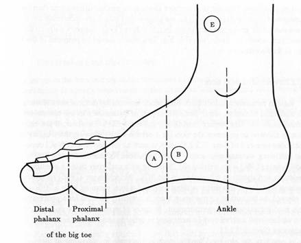
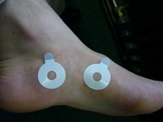
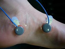
Figure 1. Medial side of the right foot with the recording sites A and B for EDA measurement.
Recording electrodermal activity from plantar sites
Recording sites
If both hands are used such as during computer work, EDA can be recorded from medial sites of the plantar (Boucsein, 1992, Fig. 28); a position recommend by Edelberg (1967). One site is above the abductor hallucis muscle (the extensor of the big toe’s base joint) adjacent to the foot sole; the other site is midway between the proximal phalanx of the big toe a point directly beneath the ankle (Fig. 1). The advantage of this site in comparison to the soles of the feet is that normal slippers or sandals can be worn during recording.
Standard procedure (as used by Boucsein & Thum 1996)
Standard electrodes (13 mm outside diameter) consist of a 8 mm contact area, covered with a sintered silver/silver chloride (Ag/AgCl) layer and a plastic rim.
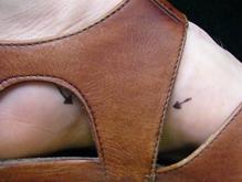
1. Recording sites should be marked first, while wearing sandals without socks, to prevent sandals from exceeding pressure on the electrodes.
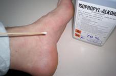
2. If recording sites are wet because of sweaty skin, they should be cleaned with 70% solution of ethanol first, to improve the adhesiveness of the EDA electrodes.
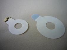
3. To improve adhesiveness, an additional bigger double-sided adhesive collar (40 mm outside/15 mm inside) may be used.
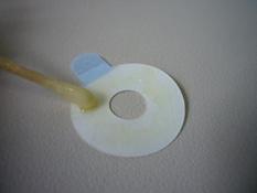
4. First, a skin friendly glue (Mastix) is brought with a Q-Tip to the adhesive site of the big collar. The glue needs 5-10 minutes to dry prior to attachment. (It becomes thread-like by touch.)
5. Thereafter, the big double-sided adhesive collar is attached to the previously marked skin sites (A, B) with its glue side.

6. The electrode is prepared as usual with a smaller size double-sided adhesive collar (20 mm outside/8 mm inside).
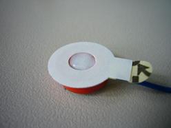
7. Then the electrode chamber is filled with isotonic electrode jelly. During this process, the protective cover of the adhesive collar still remains.
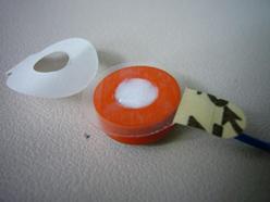
8. Remove the surplus jelly from the cover of the collar with a little paper. Then the protective cover can be removed.
9. Place the electrodes with the small collars centered on the big collars, so that the electrode paste is in contact with the skin. Mastix may be used for additionally gluing the two collars together (not shown).

10. Additionally, the collars are fixed with a skin friendly adhesive tape (Transpore) on the left and right side of the electrodes.
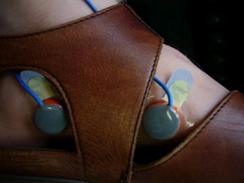
11. Put on the sandals. Socks can also be worn. Care should be taken that the electrodes are not under pressure and fit to the spare area of the sandal.
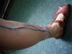
12. Tape is also used for fixation of the electrode cables to avoid strain on the electrodes.
Literature:
Boucsein, W. (1992). Electrodermal activity. Plenum Press: New York.
Boucsein, W. & Thum, M. (1996). Multivariate psychophysiological analysis of stress-strain processes under different break schedules during computer work. In J. Fahrenberg & M. Myrtek (Eds.), Ambulatory assessment; computer-assisted psychological and psychophysiological methods in monitoring and field studies (305-313). Seattle: Hogrefe & Huber Publishers.
Edelberg, R. (1967). Electrical properties of the skin. In C. C. Brown (Ed.), Methods in psychophysiology (1-53). Baltimore: Williams & Wilkins.